Signalment, History, and Physical Exam Findings
Just prior to opening our Rockville location, in July of 2014, I received a call from a local surgeon, Dr. Peter Lotsikas at Skylos Sports Medicine. Our two companies work closely together. As is par for the course with Dr. Lotsikas, he always seems to have something obscure for us to image.
 Dr. Lotsikas described Missey’s history, stating that she had been lame for about
5 months (beginning acutely in January 2014 when she was at the beach with her family). She had been on several courses of anti-inflammatories and had been to see another specialist and a rehabilitation facility. Despite these therapies, Missey had continued to decline and the owners elected to bring Missey to him and get his opinion.
On Physical Examination, Dr. Lotsikas noted that Missey was Grade III/V lame on her right front leg, and she would off-load her leg when sitting. She also had significant spasming of the limb when he abducted or extended the shoulder and there was moderate muscle atrophy of the shoulder. Her elbows palpated normally as did her cervical spine. Missey’s labwork and radiographs were unremarkable. Dr. Lotsikas was concerned that Missey either had a severe shoulder injury or possibly a herniated disk in her neck. He asked if we could image both areas on Missey. My response was, “Of course we can! You know that. Send her over and we’ll figure out what is wrong.” Then he said, “Oh, but I forgot to tell you, Missey is a 3.5 kg, 7 YO, SF Maltese.”
I paused as our normal shoulder injury patient for imaging is a large breed dog. “Well that’s a new one,” I said, “but we both know the power, utility and value that MRI provides when looking for the source of a lameness. I’m confident if there is something there to find, we will find it.”
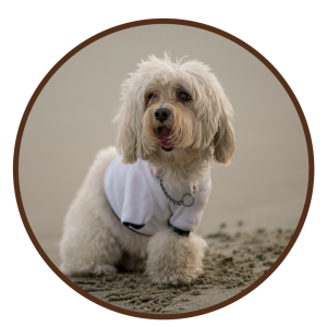
 Missey arrived at our Rockville facility on opening day. Being our first patient made her special enough, not to mention, both Missey and her parents are just about the nicest family you could ever meet. They were devastated by Missey’s condition stating that she had been in pain for 5-6 months now and they just didn’t know what else to do. They were so thankful Dr. Lotsikas had sent her in for an MRI.
Diagnostics
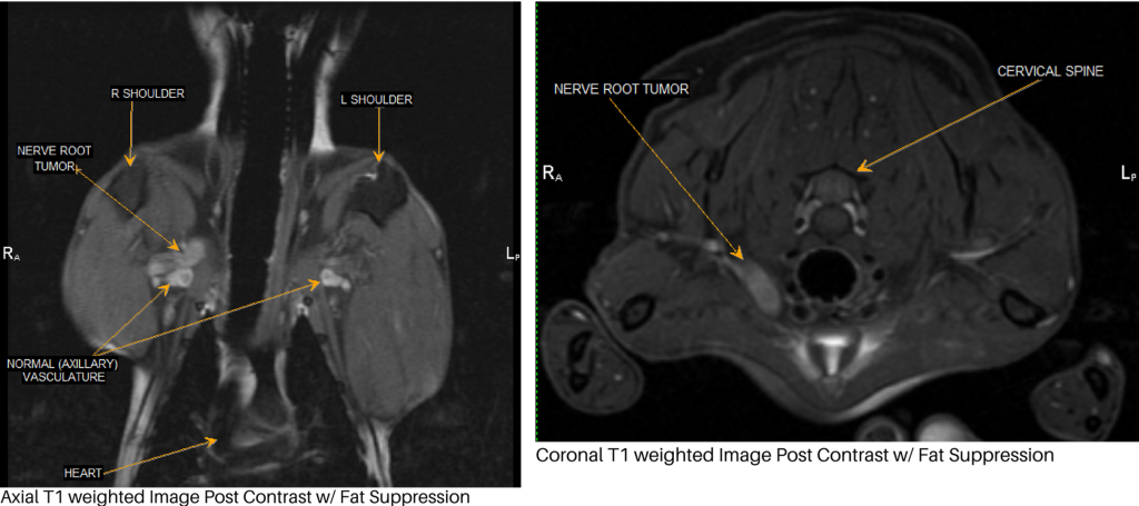
Missey’s images revealed a Right sided Peripheral Nerve Sheath Tumor (PNST) which fully explained her clinical signs. Also, note on the image on the right there is significant muscle atrophy of the affected shoulder. While these tumors are uncommon, it should be a consideration in patients that are lame with a significant degree of muscle atrophy.
Interestingly, at BAVI we just imaged another Maltese with a Right sided PNST in the same exact location.
Treatment and Follow-Up
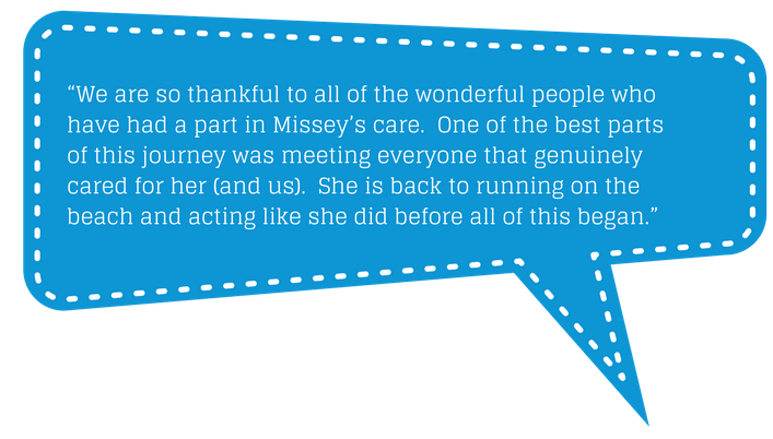
Missey went back to Dr. Lotsikas for a Right thoracic limb amputation followed by IMRT (Intensity Modulated Radiation Therapy) with Dr. Ira Gordon of The Oncology Service in Leesburg, VA. We serially followed Missey every 6 months with MRI. At 1 year, her owners felt that she was not acting herself and painful again. Indeed, they were correct, as there was evidence of tumor regrowth. Missey had a follow up surgery with Dr. Lotsikas. Clean margins were obtained and Missey also had a second course of IMRT with Dr. Gordon. We have been following Missey for another year with MRI and are happy to report that she is doing wonderfully with no obvious evidence of tumor regrowth over 2 years after her initial diagnosis.
Click HERE for a PDF version of this case study.
Learn More about MRI
Technology is changing the way we practice medicine, but it can be very difficult keeping up with all of the advances. Let us help. We are available to meet with you and your staff to discuss the value of MRI and CT imaging in the practice of veterinary medicine. Call us today to arrange a meeting. We’d be happy to bring in breakfast, lunch, or dinner.